Tendons are the strap-like elastic structures that attach muscles to the bones on which they act. Most tendons are relatively short and are rarely damaged. However, the long tendons of the limbs are vulnerable to damage during exercise or as a result of trauma. The flexor tendons are the most important long tendon structures prone to injury.
Where are the flexor tendons located?
The flexor tendons, which are made up of the deep digital flexor tendon (DDFT) and the superficial digital flexor tendon (SDFT), run down the back of the limb from the level of the knee/hock down to the foot. The SDFT ends on the pastern and the DDFT ends on the back of the pedal bone. At the level of the knee/hock, along with the fetlock and pastern region, the tendons are enclosed by a fluid filled sheath. The most commonly recognised sheath is the digital sheath at the fetlock/pastern region, with the sheath at the hock called the tarsal sheath and the sheath at the knee called the carpal sheath. Several strong, short annular ligaments help to keep the tendons in place in the regions of high movement such as joints.
The tendons themselves are composed of longitudinally arranged bundles of fibres. The blood supply to tendons and ligaments are poor compared to muscles and other tissues. This results in poorer healing of any injury.
What are the different types of tendon injuries?
Injury to these tendons occurs most commonly during exercise. Strenuous exercise can result in tearing of fibres, especially in unfit horses, or in horses which are over stretching tendons in fast work, on unlevel ground or during jumping at speed. The degree of damage to the tendons can range from minor, with minimal fibre damage, to severe with total tendon rupture.
Most frequently, a proportion of fibres are damaged in a localised area within the tendon called a zone. This may form a discrete hole which extends for a variable length of the tendon.
A knock to a tendon may result in slight bruising or could lead to severe damage leading to total tendon rupture. Sharp trauma, which cuts through the skin, can vary from minor tendon damage to partial or full thickness laceration of the tendon. If a tendon sheath is involved, these can lead to a potentially life-threatening infection if not dealt with promptly.
What are the first signs of tendon injury?
Damage to a tendon usually results in inflammation which we commonly feel as heat and swelling. Minor fibre damage leads to slight enlargement of the affected part of the tendon which feels warmer than the corresponding area of the opposite limb. Mild tendon sprains often do not cause lameness. If there is severe tendon damage, the limb can become very painful, with the toe tipped upwards or the fetlock may sink at the walk. In cases of tendon sheath sepsis the horse will also be very lame at walk.
How do we diagnose tendon injuries?
If you suspect that your horse has a tendon injury, you should call your vet for advice and an appointment. A clinical examination will help to confirm or alleviate concerns by looking for heat and pain on palpation. The extent of damage by look and palpation is difficult to assess accurately and an ultrasound scan approximately one week after injury will allows the vet to visualise and asses the extent of the damaged structure(s) if they are above the hoof capsule.
What are the treatment options?
There are several different treatment options for tendon injuries, none of which provide a guaranteed permanent return to soundness. Damaged tendon heals with irregularly arranged fibres and scar tissue. These tendons are less elastic than the original structure, thus the repair is weaker and more prone to re-injury than a healthy tendon.
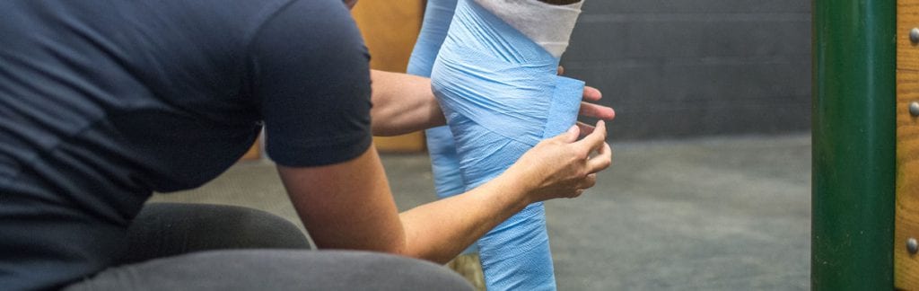
Initial treatment in the 10-14 days after an injury usually involves:
Box rest, with;
- Ice application or cold hosing two to three times daily and/or application of kaolin poultice.
- Bandaging to immobilise the limb.
- Anti-inflammatories such as ‘Bute’ to aid in reduction of swelling and provide pain relief.
These steps are aimed at reducing the initial inflammation and pain along with preventing any further injury. Once the initial inflammation has stabilised and confirmation of the severity of injury has been established, a controlled exercise program can be started.
Controlled exercise
This is the most important aspect of recuperation and treatment. Your vet will advise you on what is best for a particular injury, but will generally start with in-hand walking whilst still on box rest with gradual increases every 1 to 2 weeks for a period of three months. In certain cases, an initial period of total box rest is advised. Horses are often out of work for six to 12 months with tendon injuries, depending on the extent of the damage. Controlled exercise helps the new tendon fibres align longitudinally and ultimately results in increased strength and flexibility of the repaired tendon.
Repeat ultrasound scans are invaluable as an aid to determining the healing process, and aid in adjusting the exercise program accordingly, such as when to introduce trot work or steady canter exercise. In certain cases, the horse will not be able to return to full competition and may require a less stressful job to reduce the risk of re-injury.
Other treatments options available
None of the treatments available reduce the horse’s lay off period, but their aim is to improve the quality of repair and reduce the risk of re-injury on return to exercise.
Tendon injections
A group of drugs called PSGAG’s can be used successfully to inject tendon injuries and assist in short term healing. More commonly, stem cells or platelet rich plasma are used, both of which are injected directly into the tendon soon after injury.
Platelet rich plasma
Platelets are little blood cells responsible for clotting blood and they contain many growth factors. When injected into a tendon, the growth factors encourage new blood vessels to grow into the injury site and for more normal tendon fibres to develop. The horses’ blood can be taken and the platelet fraction harvested and injected immediately into the tendon. This is done under light sedation with a nerve block to de-sensitise the limb.
Stem cells
Stem cells live in the bone marrow and have the ability to grow into many different types of tissue, according to where they find themselves. Bone marrow also contains lots of growth factors, much like platelets. Bone marrow can be aspirated from the horse’s pelvis or sternum and sent to a specialist lab to culture more stem cells. These are then injected into the injured tendon along with growth factors in the liquid they are suspended in. The stem cells then turn into new tendon cells (a process that happens very poorly normally) providing a better quality of tendon repair.
Surgery
Surgery is sometimes performed in the case of tendon lacerations to help oppose the edges of the damaged tendon, or in the case of tendon sheath sepsis, to remove infection. Both of these procedures are carried out under general anaesthesia.
In certain cases, the accessory ligament of the SDFT is cut to allow the muscle and tendon to stretch further and reduces the incidence of SDFT re-injury. Again, this needs to be performed under general anaesthesia. However, this is not currently routinely performed as it can increase the risk of a suspensory ligament injury after surgery.
- Advice published on horsehealthprogramme.co.uk